Fluorescein and Indocyanine angiography for global appraisal of posterior uveitis, 螢光素與吲哚菁綠血管攝影於後葡萄膜炎的全面評估
後葡萄膜炎常波及視網膜與深層脈絡膜。透過螢光眼底血管造影與靛青綠血管造影的雙重檢查,能完整評估淺層血管與深部病灶發炎程度,協助醫師精準調整用藥與追蹤療效。

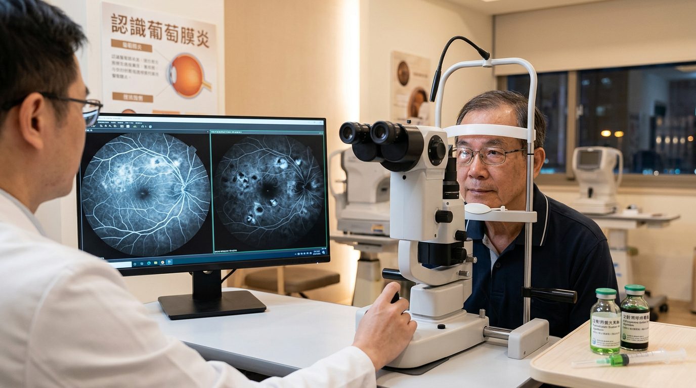
2026Oct 歐洲視網膜學會, 維也納, 黃奕修教授與會心得
Carl P. Herbort Jr, 演講 Fluorescein and Indocyanine angiography for global appraisal of posterior uveitis, 螢光素與吲哚菁綠血管攝影於後葡萄膜炎的全面評估
Herbort 教授主張 雙重血管造影 是評估後葡萄膜炎不可或缺的黃金標準
多年來,Carl P. Herbort Jr. 教授及其研究團隊的核心結論指出,螢光眼底血管造影(FA)與靛青綠血管造影(ICGA)具有高度互補性,兩者聯合使用(Dual FA/ICGA)構成了全面評估與精準追蹤後葡萄膜炎的黃金標準 (De Simone et al., 2026; Herbort et al., 2023; Papasavvas & Herbort, 2023)。
雙重造影的互補性與臨床診斷
Herbort 教授強調,單靠臨床眼底檢查或單一造影技術無法完整評估後葡萄膜炎的發炎程度 (De Simone et al., 2026; Herbort, 2018)。FA 利用小分子螢光素在可見光下的特性,主要呈現視網膜血管炎、黃斑部水腫與視神經盤發炎等淺層結構改變 (Papasavvas & Herbort, 2023; Herbort et al., 2020; Chandrasekaran et al., 2022)。相比之下,ICGA 具有近紅外線波長穿透視網膜色素上皮(RPE)以及高蛋白結合率的大分子特性,能夠穿透屏障直接檢視脈絡膜毛細血管與基質層 (De Simone et al., 2026; Papasavvas & Herbort, 2023; Herbort et al., 2020)。
藉由標準化的造影檢查程序,Herbort 將脈絡膜血管病變系統性地分類為原發性發炎性脈絡膜毛細血管病變(如 MEWDS、APMPPE)與基質性脈絡膜炎(如 VKH 氏病、鳥槍彈樣脈絡膜視網膜病變及結節病) (De Simone et al., 2026; Herbort et al., 1998; Bouchenaki et al., 2002)。在諸如伏格特-小柳-原田病(VKH)或眼結節病中,脈絡膜發炎常呈現為隱匿性的暗點(hypofluorescent dark dots)或擴散性高螢光,這些病灶在臨床檢查或純 FA 影像中極易被遺漏 (De Simone et al., 2026; Herbort et al., 1998; Herbort, 2018; Wolfensberger et al., 1999)。
| 造影檢查模式 | 主要觀察解剖層次 | 典型病理特徵 | 臨床診斷價值 |
|---|---|---|---|
| 螢光血管造影 (FA) | 視網膜血管、視神經盤、淺層黃斑部 (Herbort et al., 2020) | 血管染色滲漏、微血管無灌注、黃斑部水腫 (Tuğal-Tutkun et al., 2010) | 評估視網膜血管炎與淺層發炎併發症 (Papasavvas & Herbort, 2023) |
| 靛青綠血管造影 (ICGA) | 脈絡膜毛細血管、脈絡膜全層基質 (De Simone et al., 2026; Herbort et al., 2023) | 低螢光暗點、血管模糊擴散、基質晚期高螢光 (Herbort et al., 1998; Tuğal-Tutkun et al., 2010) | 揭露隱匿性脈絡膜肉芽腫與灌注缺損 (De Simone et al., 2026; Bouchenaki et al., 2002) |
| 雙重聯合造影 (FA/ICGA) | 視網膜與脈絡膜全層結構 (Herbort et al., 2023; Papasavvas & Herbort, 2023) | 獨立評估雙層發炎程度(半定量計分系統) (Herbort et al., 2023; Tuğal-Tutkun et al., 2010) | 全面指引全身性免疫抑制劑調配與停藥 (De Simone et al., 2026; Herbort, 2018) |
Figure 1: Comparison of FA, ICGA, and dual angiography capabilities in posterior uveitis
客觀量化計分與新興影像技術的定位
傳統國際葡萄膜炎命名標準化(SUN)僅依賴主觀的玻璃體混濁度(vitreous haze)來評估後葡萄膜炎嚴重度,Herbort 指出該標準極不充分且容易忽略無玻璃體發炎的脈絡膜病變 (Herbort et al., 2023; Papasavvas & Herbort, 2023; Herbort, 2018)。為此,團隊建立了雙重血管造影半定量評分系統(FA 滿分 40 分,ICGA 滿分 20 分乘係數 2 調整),讓臨床醫師能夠獨立量化視網膜與脈絡膜的發炎活動度 (Herbort et al., 2023; Tuğal-Tutkun et al., 2010)。該客觀計分模式已被證實能有效指引類固醇與免疫抑制劑的升級或安全減量,並廣泛應用於臨床試驗評估 (De Simone et al., 2026; Herbort et al., 2023; Massy & Herbort, 2017; Sadiq et al., 2020)。
針對光學同調斷層血管造影(OCT-A)等非侵入性技術的興起,Herbort 多次撰文反駁「OCT-A 可完全取代染劑血管造影」的觀點 (Herbort et al., 2023; Herbort et al., 2022)。他指出 OCT-A 無法動態呈現發炎滲漏(leakage),且受限於視野狹窄、易產生假影,難以有效監控涉及全眼底或深部基質的廣泛性發炎 (Herbort et al., 2023; Herbort et al., 2022)。
Herbort 教授展示了 FA、ICGA 與非侵入性工具在評估脈絡膜毛細血管無灌注病變時的互補表現。 Figure 2: Multimodal imaging demonstrating nonperfusion dynamics across ICGA, BAF, and OCT modalities (Herbort et al., 2023)總結而言,Herbort 教授數十年來的研究結論確立:雖然非侵入性影像技術(如 EDI-OCT、OCT-A)是極佳的輔助工具,但雙重 FA/ICGA 仍是掌握後葡萄膜炎發炎本質、指導精準免疫治療及避免不可逆視力損傷時無法被完全取代的基石 (De Simone et al., 2026; Herbort et al., 2023)。
References
Bouchenaki, N., Cimino, L., Auer, C., Tran, V., & Herbort, C. (2002). Assessment and classification of choroidal vasculitis in posterior uveitis using indocyanine green angiography.. Klinische Monatsblatter fur Augenheilkunde, 219 4, 243-9. https://doi.org/10.1055/s-2002-30661
Chandrasekaran, P., Aljneibi, S. H., Agarwal, A., Pichi, F., & Neri, P. (2022). Interpreting posterior uveitis by integrating indocyanine green angiography, optical coherence tomography, and optical coherence tomography angiography data: A narrative review. Saudi Journal of Ophthalmology, 36, 344 - 355. https://doi.org/10.4103/sjopt.sjopt_69_22
De Simone, L., Byszewska, A., AlBloushi, A., Genovese, R., Papasavvas, I., & Herbort, C. J. P. (2026). Indocyanine green angiography in uveitis. Saudi Journal of Ophthalmology, 40, 199 - 228. https://doi.org/10.4103/sjopt.sjopt_225_26
Herbort, C., Takeuchi, M., Papasavvas, I., Tuğal-Tutkun, İ., Hedayatfar, A., Usui, Y., Ozdal, P., & Urzua, C. A. (2023). Optical Coherence Tomography Angiography (OCT-A) in Uveitis: A Literature Review and a Reassessment of Its Real Role. Diagnostics, 13. https://doi.org/10.3390/diagnostics13040601
Herbort, C. (2018). Comparison of Retinal and Choroidal Involvement in Sarcoidosis-related Chorioretinitis Using Fluorescein and Indocyanine Green Angiography. Journal of Ophthalmic & Vision Research, 13, 426 - 432. https://doi.org/10.4103/jovr.jovr_201_17
Herbort, C. J. P., Tuğal-Tutkun, İ., Mantovani, A., Neri, P., Khairallah, M., & Papasavvas, I. (2020). Advances and potential new developments in imaging techniques for posterior uveitis Part 2: invasive imaging methods. Eye, 35, 52 - 73. https://doi.org/10.1038/s41433-020-1072-0
Herbort, C., Lehoang, P., & Guex-Crosier, Y. (1998). Schematic interpretation of indocyanine green angiography in posterior uveitis using a standard angiographic protocol.. Ophthalmology, 105 3, 432-40. https://doi.org/10.1016/s0161-6420(98)93024-x
Herbort, C., Papasavvas, I., & Tuğal-Tutkun, İ. (2022). Benefits and Limitations of OCT-A in the Diagnosis and Follow-Up of Posterior Intraocular Inflammation in Current Clinical Practice: A Valuable Tool or a Deceiver?. Diagnostics, 12. https://doi.org/10.3390/diagnostics12102384
Massy, R., & Herbort, C. (2017). Contribution of Dual Fluorescein and Indocyanine Green Angiography to the Appraisal of Presumed Tuberculous Chorioretinitis in a Non-endemic Area. Journal of Ophthalmic & Vision Research, 12, 30 - 38. https://doi.org/10.4103/2008-322x.200157
Papasavvas, I., & Herbort, C. P. (2023). Review on imaging methods in non-infectious posterior uveitis, principles, relevance, and practical clinical applications to disease entities. Oftalmologicheskii Zhurnal. https://doi.org/10.31288/oftalmolzh20233959
Sadiq, M., Hassan, M., Afridi, R., Halim, M. S., D., Sepah, Y., & Nguyen, Q. (2020). Posterior segment inflammatory outcomes assessed using fluorescein angiography in the STOP-UVEITIS study. International Journal of Retina and Vitreous, 6. https://doi.org/10.1186/s40942-020-00245-w
Tuğal-Tutkun, İ., Herbort, C., & Khairallah, M. (2010). Scoring of dual fluorescein and ICG inflammatory angiographic signs for the grading of posterior segment inflammation (dual fluorescein and ICG angiographic scoring system for uveitis). International Ophthalmology, 30, 539-552. https://doi.org/10.1007/s10792-008-9263-x
Wolfensberger, T., Piguet, B., & Herbort, C. (1999). Indocyanine green angiographic features in tuberculous chorioretinitis.. American journal of ophthalmology, 127 3, 350-3. https://doi.org/10.1016/s0002-9394(98)00325-0
本文相關的疾病專區
相關內容
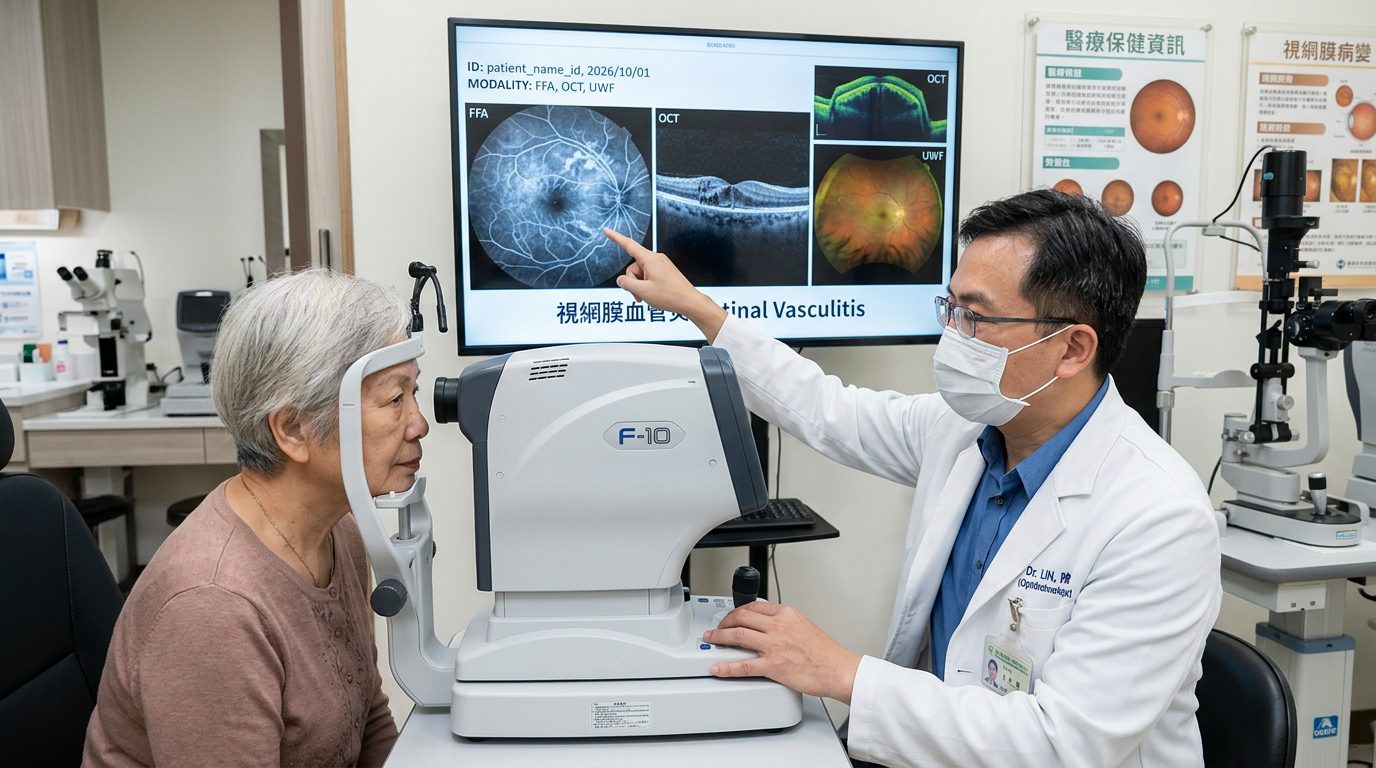
2026/Oct Euretina Vienna, 心得 Retinal vasculitis 視網膜血管炎:全球文獻聚焦於視網膜血管炎的標準化定義與多模態影像評估
視網膜血管炎是嚴重的眼內發炎反應。最新國際共識建立了標準化診斷名詞,並強調螢光眼底血管造影結合多模態影像的必要性,早期搭配全身性免疫調節或生物製劑能有效保護視力。

葡萄膜炎分類: 依發病位置分類
依前、中、後段與全葡萄膜炎介紹SUN解剖位置分類系統。
什麼葡萄膜炎是台灣最常見?
台灣最常見的葡萄膜炎類型為前葡萄膜炎(虹彩炎),在已知特定病因中,以 HLA-B27 相關葡萄膜炎最為常見,感染性則以疱疹病毒引起者較多。